

Recognition, hyaluronidase treatment, and the evidence for ultrasound-guided management
A clinical review drawn from fifteen source publications
Contents
- You Notice the Skin Turns White
- The Natural History of an Untreated Filler Vascular Occlusion
- What a Filler-Induced Vascular Occlusion Is
- Compression is no longer accepted
- Embolism: the best-evidenced mechanism
- Vasospasm: back, in a different role
- Microembolism: what the artificial vessel model shows
- Rheology and cannula gauge predict the behaviour
- The thrombotic component, and a newer therapeutic idea
- The three models are not mutually exclusive
- How Often Filler Vascular Occlusion Happens
- Why Timing Is the Whole Game
- The two causes of permanent tissue deficit
- The History: From Fixed Daily Doses to Pulsed Flooding
- The 2017 high-dose pulsed protocol
- What the pooled evidence says about hyaluronidase dose
- Adjuncts: what works, what does not
- Treating an Occlusion Caused by a Non-HA Filler
- Where Ultrasound Is Taking Us
- The caveats, stated plainly
- Bringing It Together: What To Do At The Chair
- Source Publications
You Notice the Skin Turns White
It is the last patient of the day. The nasolabial fold has been treated without incident, and then, somewhere in the middle of the second pass, the skin over the alar base goes pale. Not bruised. Not swollen. White, in a patch that does not match the surrounding cheek, and it is not pinkening back up the way a transient vasoconstrictive blanching should. You ask the patient how it feels. She says it stings, more than she expected. The sun is going down, you have one set of hands in the room, and the next sixty minutes will determine whether this patient leaves your clinic with her own skin.
Every injector has rehearsed this in theory and almost nobody has rehearsed it in practice. That gap is what this article is about. The window between the moment the skin blanches and the moment a blister forms is roughly seventy-two hours, and how that window is used is the difference between a patient who walks out with a bruise and a patient who walks out with a scar, a depressed contour that no amount of later filler will fully correct, or, in the worst case, an eye. What follows is what the evidence says to do, drawn from twelve publications spanning a century of observations on the problem, the 2017 high-dose pulsed protocol that remains the standard of care, comparative and meta-analytic evidence on dosing and timing, case series on non-HA fillers, and the ultrasound literature that has begun to reframe what a vascular occlusion actually is.
The recurring message across all of it is that recognition is clinical, treatment is time-critical, and the direction of travel is toward smaller doses given more precisely. Before any of that, though, the clinician in that room needs to know exactly what they are watching happen to the skin in front of them.
The Natural History of an Untreated Filler Vascular Occlusion
The untreated course is well mapped, and it is the reference against which any treatment decision is made. The clinical staging below, from the Murray guideline as reproduced in the Soares review, is consistent with the milestones described across the rest of this literature.
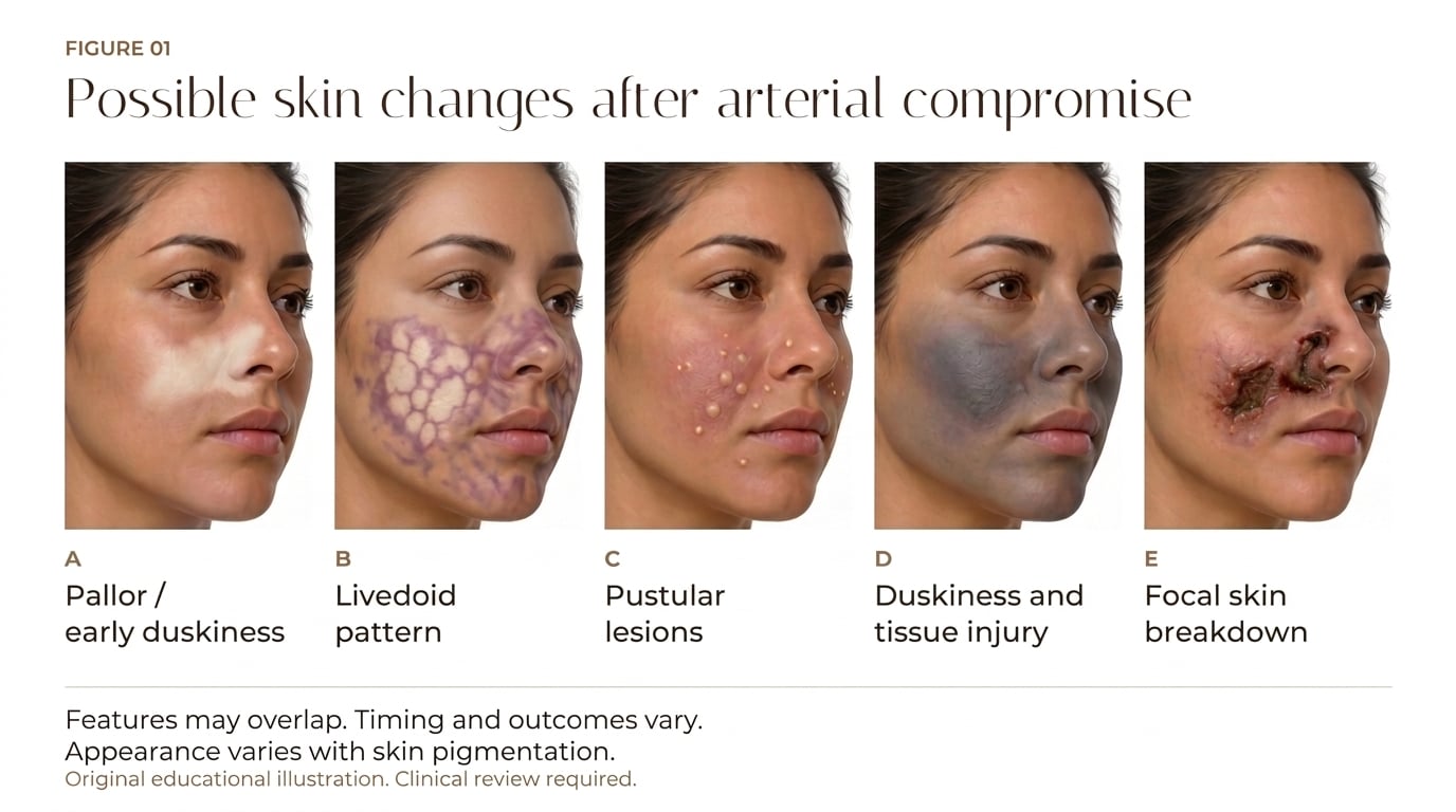
Table 1. Untreated progression of a vascular occlusion and what each stage implies
Swipe or scroll horizontally to read all columns. The first column stays visible.
| Stage | When | What is seen | What it means |
|---|---|---|---|
| Blanching | Seconds | A momentary white patch over the injected area, often missed | Fully reversible; a single dose of enzyme usually restores normal skin with no residue |
| Livedo reticularis | Up to 72 h | Net-like violaceous mottling; capillary refill markedly slowed | The signature sign. Looks stable, which is what makes it dangerous |
| Barrier breakdown | Days 3 to 5 | Partial desquamation with rising bacterial bioburden | The skin barrier is failing; infection risk now compounds the ischemic injury |
| Coagulative necrosis | Days 5 to 10 | Dark dry crust over dead skin | Frank skin death; healing will now be prolonged |
| Eschar | Weeks | A leathery crust that may persist for several weeks | Scarring, and in patients of colour often persistent dyschromia |
| Visual loss, if ocular | Immediate | Ocular pain, ptosis, ophthalmoplegia | Typically permanent; the retina tolerates only about 90 minutes of ischemia |
Two details in that sequence deserve emphasis because they explain why clinicians get caught out. Livedo reticularis is not merely a colour change: it represents deoxygenated blood pooling in the post-capillary venules of the skin because arterial supply has been blocked upstream. It looks like a stable rash and it is not. The tissue at that stage is already ischemic and already dying at a rate determined by its metabolic demand. By the time the barrier begins to fail, the skin has been without perfusion for days, bacteria are colonising compromised tissue, and the window for a scar-free outcome has closed.
Untreated, the process also changes shape. Partial obstruction that is bypassed by collaterals may produce no ischemia at first, and then declare itself hours later when the material moves downstream and blocks those collaterals. This explains the intimidating phenomenon of delayed-onset vascular occlusion, where a patient is entirely normal at the end of treatment and frankly ischemic an hour later. Pooled case data put the mean interval from injection to symptom onset across reported events at around 56 hours, though the variability is wide. It also explains why symptoms do not always appear where you injected: in 15% of cases in one series, skin changes surfaced in an adjacent territory, with nasolabial fold injections presenting on the nose and nose injections presenting at the glabella, because one perforator territory can border or overlap another.
Disproportionate pain and skin discolouration are the two findings that should stop you: they are present in roughly 80% and 70% of confirmed ischemic skin injuries respectively. Pain often precedes the visible change, because it emerges as the included lidocaine is metabolised and acidosis builds in the underperfused tissue.
What a Filler-Induced Vascular Occlusion Is
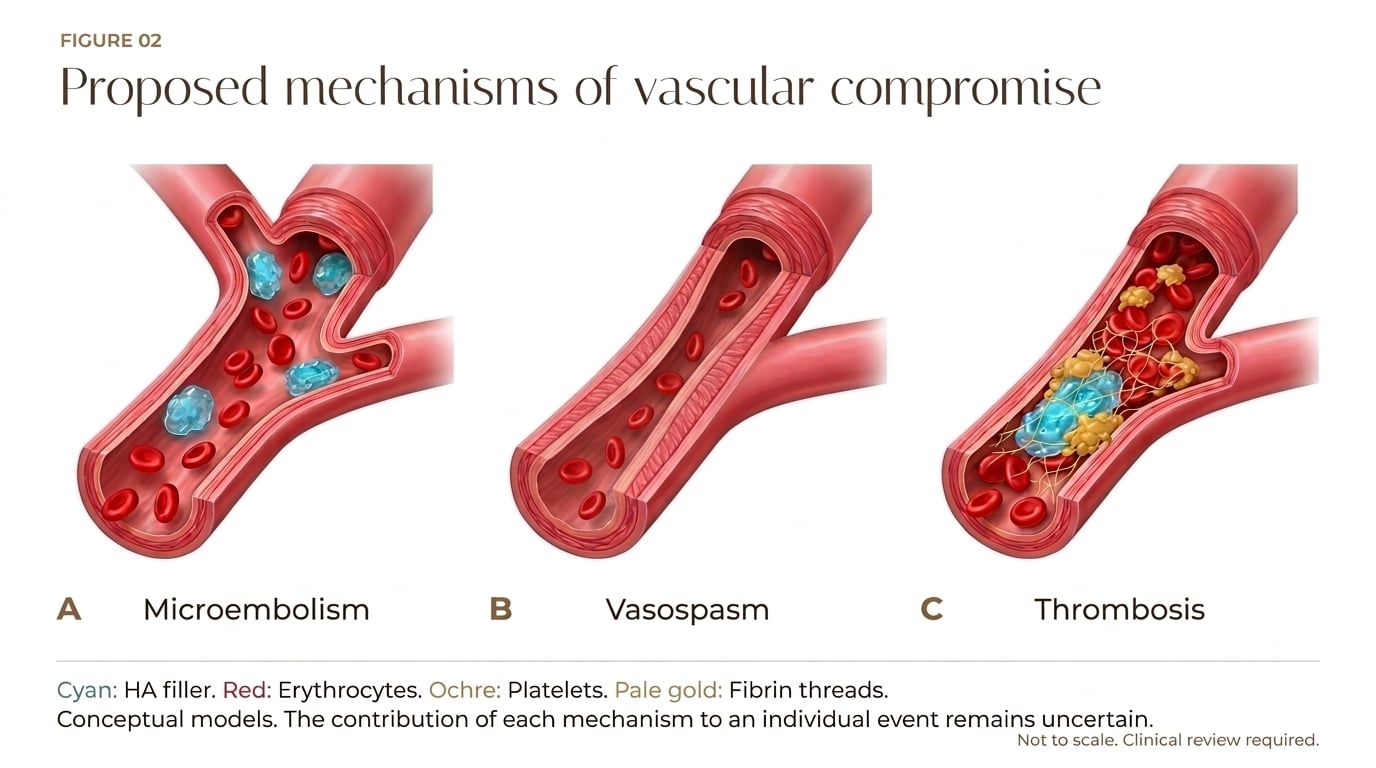
For years, teaching offered three competing explanations for why a filler injection kills skin. A filler embolus could mechanically block an artery. An external deposit could compress a vessel, the garden-hose analogy. Or the vessel wall could go into spasm. Only two of those have survived, and the one that did not is the one that used to be taught most confidently.
Compression is no longer accepted
The decisive evidence is experimental. In mouse and rabbit models of filler-induced ischemia, arterial compression by filler could not be induced and did not produce any prolonged period of significant underperfusion or ischemia. You cannot squeeze a facial artery shut with a bolus of gel and keep it shut. The anatomy argues the same way: single-point obstructions of facial vessels are easily overcome by a generous redundant supply from adjacent angiosomes through true anastomoses, which dilate after filler placement, and even bilateral ligation of the external carotid arteries historically performed for intractable epistaxis was survivable without tissue necrosis because the internal carotid system compensated. Compression is now recognised as a valid alternative mechanism only in narrow circumstances: distal arterial branches in previously operated tissue with a tenuous blood supply, or tissue nourished by end-arteries with no cross-perfusion at all.
Embolism: the best-evidenced mechanism
Histopathology of ischemic tissue shows filler sitting in the lumen of arteries, which is direct, visible proof. The volume of the named vessels reinforces how little it takes. The average intra-arterial volume of the supraorbital, supratrochlear and dorsal nasal arteries approaches 0.1 mL, so a bolus of a fraction of a millilitre into the wrong place can fill those vessels and travel retrograde into the proximal ophthalmic artery and beyond. There is also a plausible route of entry that has nothing to do with a needle tip sitting inside a lumen. If a vessel is penetrated by a needle or cannula, filler deposited even some distance beyond the vessel can backtrack along the needle track and enter the vessel, following the path of least resistance. In scarred tissue from previous trauma or a long series of earlier filler treatments, that track becomes a conduit. This is also why aspiration before injection can mislead: bright red blood is informative, but a negative result proves nothing.
The rheology of the gels matters here. Modern fillers are crosslinked hyaluronan with a degree of modification between 1 and 10%, molecular weights of 100 to 600 kDa, and HA concentrations of 15 to 24 mg/mL, giving them viscoelastic solid behaviour that resists shear and compressive deformation. The pressures required to make them flow smoothly through a needle are well above mean arterial pressure: one study found that an average injection pressure of 166 mmHg was enough to inoculate the supratrochlear artery with a common filler and retrogradely fill the ophthalmic artery back to the takeoff of the retinal artery, while another showed even soft fillers with low elastic modulus extruding from a 25-gauge needle at 580 mmHg. The clinical translation of that physics is the familiar rule: inject low and slow, in small boluses, and never chase a bleb with force.
Vasospasm: back, in a different role
Vasospasm was historically dismissed because HA is deliberately innocuous, chosen precisely because it does not irritate tissue, and because mechanical vessel irritation usually causes only transient spasm. It has since returned to the discussion in a form that fits the ultrasound evidence. In every one of 39 confirmed events imaged with Doppler in a comparative case series, the pattern was consistent: hypervascularity around the main arterial trunk, with the filler sitting as a hypoechoic to isoechoic mass in the perivascular space adjacent to or surrounding that vessel, while the superficial tissue showing the visible skin changes had little to no vascular signal. Critically, no filler was found in the superficial tissue where the necrosis was declaring itself. The obstruction was deep; the skin was only the downstream witness.
That observation is hard to square with a simple embolus model, and it underpins the perforasome concept, adapted from Taylor's angiosome theory. A single perforating artery supplies a discrete territory of soft tissue, with neighbouring territories connected by anastomoses that come in two functional types. True anastomoses are permanent wide connections that enlarge the accessible arterial territory and can carry filler from one angiosome into another. Choke vessels act as gatekeepers, restricting flow between adjacent territories, and they may reflexively constrict in response to a filler insult, which limits the spread of filler to the neighbouring angiosome but also cuts off potentially vital collateral perfusion to the threatened tissue. That double-edged behaviour is part of why occlusion patterns vary so much between patients.
If filler injection triggers vasospasm of a perforator and its anastomoses, the whole perforasome goes ischemic, which is why the visible skin pattern is often geometric or wedge-shaped rather than random, and why it can map onto the superficial fat compartments of the face that Rohrich and Pessa described. On this reading the enzyme works less as an antidote dissolving a plug and more as a vasodilator releasing a spasm, since hyaluronidase has independently been shown to reverse microcirculatory vasospasm in animal models. That reframing explains something the embolus model struggles with: the same enzyme resolves occlusions caused by calcium hydroxylapatite (CaHA) and poly-L-lactic acid, neither of which it can chemically degrade.
Microembolism: what the artificial vessel model shows
The most direct evidence on mechanism comes from an artificial perfusion model rather than from patients, and it answers a question the clinical literature could only infer. Five commercial HA fillers were injected as 0.2 mL boluses through 22 G and 27 G microcannulas into a branched microtubule network tapering from 1,000 microns down to 200 microns, with flow parameters matched to the paramandibular segment of the facial artery. What the model showed is that HA gels are not the static intraluminal plugs that early teaching assumed. They fragment extensively into thousands of microparticles, averaging 2,088 per trial with a mean cross-sectional area of 0.140 mm2, and those fragments disperse progressively downstream, behaving as deformable embolic suspensions rather than cohesive blocks.
Occlusion clustered sharply by channel calibre. Most products kept channels of 400 microns and above patent in more than 90% of branches, but in vessels of 300 microns or less, 88% showed some degree of blockage, and at 200 microns obstruction appeared in 90% of channels with complete occlusion in 60%. That 100 to 300 micron range is not arbitrary: it corresponds to resistance arterioles, to the arterio-arterial anastomoses of the facial subcutaneous and subdermal plexuses, and to the central retinal artery and its proximal branches. The implication is uncomfortable. Microemboli at that scale can negate collateral perfusion, converting a vascular field that is normally redundant into a functionally end-arterial one, which is a mechanistic explanation for the conserved, angio-perforasomal territory patterns that clinicians see on the skin.
Rheology and cannula gauge predict the behaviour
Not every filler embolises the same way, and the differences track the gel's rheology. Solid-character gels with low energy dissipation factors fracture in a brittle pattern, yielding ovoid microparticles with real occlusive potential. High-fluidity gels do something quite different: a soft product with an energy dissipation factor of 1.27 formed long filamentous wisps that were largely non-occlusive, tending instead to coat the vessel wall. One high-elasticity product in the series fragmented finely enough to clear even 200 micron channels, which is a reminder that elastic strength alone does not predict occlusive behaviour.
Cannula gauge matters too, and in a direction that complicates the standard advice. A 22 G cannula produced both larger particles and a higher rate of occlusion than a 27 G, 31% against 17%, and the effect was concentrated in the large-particle, high-elasticity products: one gel occluded 86% of channels when extruded through 22 G against 27% through 27 G. The mechanism is shear. Passing through a narrower conduit imposes more shear strain, and more shear means more fragmentation, which means smaller particles. So the wider, gentler device delivers a less fragmented bolus. This sits alongside the earlier population finding that cannulas carry lower occlusion odds than needles overall, and the two are not in conflict: a cannula is less likely to enter the vessel in the first place, but when it does, or when a large-bore cannula is used with a stiff, large-particle product, the resulting embolus may be cruder.
The model also showed that the vasculature itself filters the material. Comparing particle sizes against an earlier macrovascular version of the same rig, every product fragmented further in the smaller-calibre network, with reductions running from 31% to 64% depending on the gel. Smaller vessels mechanically break down what passes through them. The authors' framing of the whole problem is worth borrowing: what determines the extent of injury is the embolic load, the volume of gel relative to the volume of the affected arterial tree. Because most arterial inoculation happens over split seconds as the needle moves, a typical load is only 10 to 100 microlitres, which is negligible in a large vessel and catastrophic in a retina.
The thrombotic component, and a newer therapeutic idea
One mechanism in the current literature is not a theory about the initial insult at all but about what the insult starts. An intraluminal bolus of HA gel creates haemostasis and a direct HA-platelet interaction capable of triggering platelet aggregation. In murine models this produces an early platelet-rich white thrombus within the gel plug that extends beyond the bolus into a fibrin-rich red thrombus. The immediacy of the process points to an inflammatory trigger from the HA itself rather than anoxic endothelial injury, and the finding that platelets are necessary is confirmed by the observation that platelet-free serum shows no clot-inducing properties when mixed with filler in vitro. The clinical translation is that the obstruction a clinician faces at hour one may be larger than the volume originally injected.
That has opened a genuinely new therapeutic avenue. In rat models of HA occlusion, flap survival was significantly better in animals given dual therapy with a thrombolytic agent (urokinase or alteplase) plus hyaluronidase than with hyaluronidase alone, and the benefit held for both subcutaneous and intravenous routes, which is important because it avoids the arterial injury risk of intra-arterial work. In humans, patients with central retinal artery occlusion treated with combined intra-arterial therapy have done comparatively better than those given monotherapy, though prompt initiation still dominates every other variable. Two problems remain: the necessarily delayed presentation of most ocular cases, and the risks of the super-selective angiography itself, including iatrogenic cerebral infarction. High-dose intravenous hyaluronidase is now being explored as a faster and more accessible route, but because the plasma half-life of the enzyme is only two to three minutes, that would likely require a continuous infusion to hold therapeutic activity at a distant site of occlusion.
The three models are not mutually exclusive
- A small embolus may seed a spasm, and a spasm may in turn create the stasis that allows a thrombus to form and extend the block. What remains true is that no study has yet proven in vivo which mechanism dominates, and the ultrasound findings in favour of the perforasome model are drawn from 39 patients in a single retrospective case series.
How Often Filler Vascular Occlusion Happens
The incidence of filler-associated vascular occlusion is genuinely low, and the best available figure comes from the largest dataset on the question: 370 board-certified dermatologists reporting 1.7 million syringes of filler injected. Occlusion risk never exceeded 1 in 5,000 syringes for any filler type. For hyaluronic acid specifically it worked out at roughly 1 in 5,700 injections with a needle versus 1 in 35,000 with a cannula, and multivariate analysis found injections with a cannula carried 77.1% lower odds of occlusion than needle injections.
Table 2. Reported incidence of filler-associated vascular occlusion
Swipe or scroll horizontally to read all columns. The first column stays visible.
| Measure | Value | Source |
|---|---|---|
| Vascular compromise, general estimate | 0.05 to 0.2% | Pooled literature |
| Blindness | 0.001 to 0.005% | Pooled literature |
| Skin necrosis or visual loss after VO | About 0.2% | Pooled literature |
| Occlusion, needle injection | 1 in 5,700 syringes | 370 injectors reporting 1.7 million syringes |
| Occlusion, cannula injection | 1 in 35,000 syringes | Same cohort |
| Occlusions with no long-term sequelae | 85% | Same cohort |
| Injectors who had seen at least one event | 28.6% (106 of 370) | Same cohort |
| Skin injuries among reported events | About 80% | FDA review, 2015 to 2020 |
| Events occurring on the face | At least 91% | Same review |
| Reported injuries with visual sequelae | Nearly 20% | Same review |
Two contextual points matter for how these numbers land in practice. First, experience and technique are protective: injectors with more than five years of filler practice had 70.7% lower odds of occlusion than those with less, and each additional injection per week in a clinician's workload lowered the odds by about 1%. Second, no anatomical site is zero-risk, though the risk is not evenly distributed. Nasolabial folds and lips were the sites most likely to be occluded, while occlusions at the glabella carried the highest mean severity. Retrospective case series report far higher complication rates than these population figures, which reflects referral bias rather than a discrepancy: complicated cases cluster in specialist clinics.
The reporting data also show that the problem is growing in absolute terms even as the per-injection risk stays low. Filler sales rose by 700% between 2005 and 2020, with roughly 3.4 million filler procedures a year in the United States alone and HA products comprising nearly 80% of them. Against that background, the published ischemic skin injuries rose 30-fold between 2000 and 2020 and filler-induced stroke reports rose by 300%. Nearly 50 cases of filler-induced blindness were published in the three years between 2015 and 2018, which is striking against only 98 in the preceding century. That history is longer than most injectors realise: paraffin injections in the 1900s produced sudden blindness, stroke and pulmonary embolism as early as 1903, and the cause was correctly identified at the time as inadvertent intravascular dissemination into the ophthalmic, cerebral and pulmonary vasculature. The FDA ordered vascular occlusion warnings onto all filler product labelling in 2015.
Why Timing Is the Whole Game
The pooled evidence converts the timeline above into numbers. Across 231 patients with HA filler complications, complete resolution of impending skin necrosis was 100% when hyaluronidase was given within 6 hours of symptom onset, falling to roughly 87% at 6 to 24 hours and about 81% beyond 24 hours, with the confidence intervals widening sharply in the later groups as delayed presentations become more heterogeneous. A separate protocol analysis found no cases of partial or complete skin loss when treatment began within 48 hours of onset. At the other end of the range, one group successfully removed HA gel as late as 63 months post-treatment for non-vascular indications, which is a reminder that filler remains removable long after the acute window has closed; what is lost with delay is the tissue, not the filler.
The reason the numbers fall so steeply is that different tissues have wildly different tolerance for ischemia. The human retina may suffer irreversible injury within 90 minutes of occlusion and possibly sooner, reflecting its extremely high basal metabolic rate. The brain tolerates only 1.5 to 4.5 hours. Skin is the most forgiving of the three at 12 to 24 hours, which is precisely why a facial skin occlusion can look recoverable for a day and then declare itself necrotic on day three. There is also a second injury on top of the first: when circulation is restored, reperfusion itself causes harm through reactive oxygen species mediated endothelial injury, which triggers a prothrombotic state that can occlude capillaries and extend the original damage.
Two mechanisms explain the drop-off in outcome, and they compound. The first is cell death on a clock: fat cells are the most ischemia-sensitive residents of the subcutaneous layer, and once they die the deficit is structural rather than cosmetic. This is the single cause of permanent volume loss after an occlusion, a depression that no later filler fully corrects because the scaffold it would sit in is gone. The second is the treatment's own physics. Pulsed dosing exists because the enzyme is simultaneously diluted by leaking serum, diffused away down its concentration gradient, and actively deactivated by tissue anti-hyaluronidase enzymes. Hours of delay mean more oedema, more enzyme washout, and a larger ischemic zone to saturate, so the delay worsens the injury and makes the rescue harder in the same step.

The two causes of permanent tissue deficit
- Both identified causes of lasting tissue loss in this literature are iatrogenic, and both are failures of conservative management rather than aggressive management: under-treatment of ischemia, and a mismanaged abscess where infection was allowed to fester before incision and drainage.
- Hyaluronidase does break down native HA, but tissue rebalances within days and does not suffer permanent atrophy from it. The enzyme is not the risk; the wait is.
The History: From Fixed Daily Doses to Pulsed Flooding
The first major breakthrough was simply the realisation that hyaluronidase was the treatment, and that the rest of the armamentarium contributed little. Harold Pierce published a 1988 protocol that became the template for the next quarter century: hyaluronidase injected directly into the affected area at around 450 to 600 IU, repeated as a single daily treatment, alongside a package of ancillary measures including topical nitropaste, hyperbaric oxygen, and vasodilators. The results were acceptable but not clean. Some patients healed completely; others went on to blistering, crusting, mild scarring and dyschromia, and outcomes were visibly worse in patients with more facial regions involved. That pattern, of some patients resolving and others not, prompted the reasoning that not enough enzyme was being used in at least some cases, and led to the flooding hypothesis: HA-obstructed vessels need to be bathed in a high concentration of enzyme for long enough to dissolve the filler.
The 2017 high-dose pulsed protocol
Claudio DeLorenzi's High Dose Pulsed Hyaluronidase (HDPH) protocol, published in 2017, is the current standard of care. Its logic is concentration over time. Hyaluronidase must diffuse across the intact arterial wall and reach the obstructed filler at a sufficient concentration for long enough to break the HA down into fragments small enough to pass through capillaries of 5 to 10 microns. Partial hydrolysis is not sufficient: partially degraded fragments can still obstruct, and may be pushed downstream to block collateral vessels. Crosslinked fillers take hours to degrade this way, whereas non-crosslinked HA is hydrolysed almost instantaneously in vitro.
Against that, three processes constantly erode the enzyme's activity. It is diluted by serum leaking from ischemic capillaries, it diffuses away from the site of high concentration, and it is actively deactivated by naturally occurring anti-hyaluronidase enzymes. A single daily dose cannot hold a therapeutic concentration. Hence the protocol's two structural changes, which are the features that distinguish it from everything before it.
- Dose scales with ischemic surface area, not a fixed number. The clinically visible patch of ischemic skin is used as a proxy for the volume of tissue that must be bathed in enzyme, since nobody can see the intravascular filler at the bedside. More ischemic skin means more units, escalating stepwise across facial territories: roughly 450 IU for a single area, up to 1500 IU where three areas are involved.
- Doses repeat hourly until reperfusion. Treatment is titrated to capillary refill, skin colour and resolution of pain, with gentle massage after each injection to maximise contact between enzyme and filler. No ancillary treatments are used at all in the current version, beyond one aspirin daily for seven days, given to reduce platelet activity even though direct evidence for its benefit is lacking here.
The reported difference is stark. In two years and several dozen cases, DeLorenzi saw no partial or complete skin loss when the protocol started within two days of onset. Instead of six weeks of healing by secondary intention, treated patients returned to normal within about three days, with no more than a few bruises and injection-site reactions from the repeated needle passes. The framework also draws a distinction that matters for prognosis: low-volume events, defined as an embolus of 0.1 mL or less, are by far the most common in experienced hands and carry a good prognosis, while high-volume events above 0.1 mL are associated with injuries to multiple systems, including loss of vision, loss of hearing and stroke.
What the pooled evidence says about hyaluronidase dose
The clinical anchor is solid; the hyaluronidase dose numbers are less tidy than the protocol implies, and two meta-analyses point in opposite directions. Pooling 223 patients across 15 studies, complete scar resolution after hyaluronidase was 77.8% overall, and patients receiving 500 IU or less resolved at 88.1% versus 69.6% for those on more than 500 IU, though the difference did not reach significance. A later analysis of 231 patients found the reverse pattern, with more than 500 IU achieving 100% resolution versus 76.9% at lower doses. The most likely reconciliation is confounding: the sicker, longer-delayed presentations receive the larger doses, and no case-series design can strip that out. What every analysis agrees on is timing, and what the more recent review recommends is a starting dose of 150 to 300 units per affected area injected subcutaneously and repeated every 30 to 60 minutes while symptoms persist, rather than a reflex 1500 IU.
Practical readiness matters as much as the number chosen. Hyaluronidase in plasma has a half-life of only two to three minutes because of plasma glycoprotein inhibitors, and in tissue it remains active for 24 to 48 hours, though some murine work shows activity falling to nothing within three to six hours. Re-dosing every one to six hours is therefore the norm, and repeat dosing is expected rather than a sign of failure. Commercially available vials typically hold 150 to 200 units per millilitre in 1 to 2 mL single-use volumes, which is a fraction of the 500 to 1500 units per dose advocated for occlusion, so stocking decisions have to be made deliberately rather than to the manufacturer's default. Recombinant human hyaluronidase is now the favoured formulation because of its lower hypersensitivity risk and higher potency, though bovine and ovine products remain available and their use as reversal agents is off-label.
Allergy testing has been a contentious topic: historical animal-derived formulations prompted routine skin testing, but reported hypersensitivity rates in clinical practice are below 0.1%, recombinant formulations have reduced immunogenicity further, and consensus guidelines now deem testing impractical in an emergency, reserving it for patients with a known history of reaction to enzymes, bee stings or animal-derived products. Hyaluronidase is contraindicated in patients with bee or wasp venom allergy. Aspirin, corticosteroids, oestrogens, furosemide, benzodiazepines and antihistamines may blunt the response and warrant larger or repeated dosing. In the instance of blindness from periocular embolism, retrobulbar injection of 150 to 200 units in 2 to 4 mL of diluent into the inferolateral orbit may be considered, although treatment is rarely successful, in part because diffusion across the arterial wall in the retro-orbital compartment is impaired.
Adjuncts: what works, what does not
A striking feature of the modern literature is how little of the traditional adjunctive package survives scrutiny. Topical nitroglycerin and oral phosphodiesterase inhibitors were conceived as vasodilators, but nitroglycerin failed to show benefit in controlled rabbit-ear studies and may have a deleterious congestive effect in affected tissue. Antiplatelet therapy with aspirin or clopidogrel has no direct evidence of benefit in occlusion, although it remains a defensible intervention by analogy with other arterio-occlusive conditions, and several modern protocols include a short course. Warm compresses, recumbent positioning and massage lack supporting evidence but carry minimal risk. Hyperbaric oxygen has never been shown objectively to improve tissue survival after filler occlusion, though it is supported by a long history of benefit in flap surgery and is associated with favourable outcomes in case reports; if used at all it belongs as an adjuvant alongside reversal therapy, started early and continued across several sessions in the first week. Reversal therapy with hyaluronidase, with or without thrombolytics, remains the first-line intervention with the greatest potential impact.
Treating an Occlusion Caused by a Non-HA Filler
The absence of an antidote for non-HA fillers was long regarded as the hardest problem in the field, and it is where the mechanism debate became clinically decisive. A 2024 multicentre case series describes three events, two after CaHA and one after poly-L-lactic acid, all resolved without necrosis or scarring using ultrasound-guided hyaluronidase injections. The CaHA cases had previously been treated with sodium thiosulphate as the primary agent, on the assumption that it dissolves CaHA, with hyaluronidase added only in the belief that it might increase tissue permeability and disperse the product. Doubts about how well sodium thiosulphate actually dissolves CaHA, combined with the observation that blood flow improved on Doppler in every case, led the group to invert the approach: hyaluronidase became the primary treatment for non-HA events, with sodium thiosulphate held in reserve.
The clinical pattern in those cases is worth knowing. The first patient presented three days after a 0.2 mL bolus of CaHA at the caudal chin with a reticulated livedoid pattern reaching from the chin toward the lower lip and down into the neck, with pain and a haematoma, and 150 units of hyaluronidase injected at the level of the SMAS restored perforator flow within minutes. The second developed bruising, swelling and a marbling pattern under the right zygomatic arch after hyperdiluted CaHA, and 300 units restored capillary refill, with a further 150 units on day 3; all symptoms resolved by day 11. The third developed blanching and delayed capillary refill in the right zygomatic area during a poly-L-lactic acid treatment, and 500 units restored normal flow in the transverse facial artery within minutes. CaHA events carry more inflammation than HA, so oral prednisolone and aspirin were added. The case-series authors' conclusion is the mechanism argument in clinical form: relaxation of the perforasomes, rather than dissolving the filler material, is what resolves the symptoms.
Where Ultrasound Is Taking Us
There is a mechanistic case for flooding that deserves stating alongside the ultrasound evidence, because the two are often framed as rivals when they are better understood as describing different parts of the same problem. If a bolus fragments and disperses into the distal microcirculation, as the perfusion model demonstrates, then the microvasculature itself becomes a degradative sink: thousands of small particles present a very large surface area to enzymatic action, mechanical stress at branch points adds to their breakdown, and diffusion distances across leaky, thin-walled capillary beds are short. On that reasoning, infiltrating the affected skin and subcutaneous tissue with hyaluronidase gives broad enzymatic exposure to precisely where the fragments have ended up, and the extravascular enzyme may also act as a reservoir, retaining activity in peri- and perivascular tissues far longer than its two-to-three-minute intravascular half-life allows. One review even argues that this mechanism is best explained by the perforasome concept rather than by DeLorenzi's diffusion model. What the flooding protocol lacks is visual confirmation that the enzyme has reached the obstruction; what targeted injection lacks is reach across a dispersed field of microparticles. Both the regional flooding and targeted-injection rationales are coherent, and the empirical question of which performs better has not been settled.
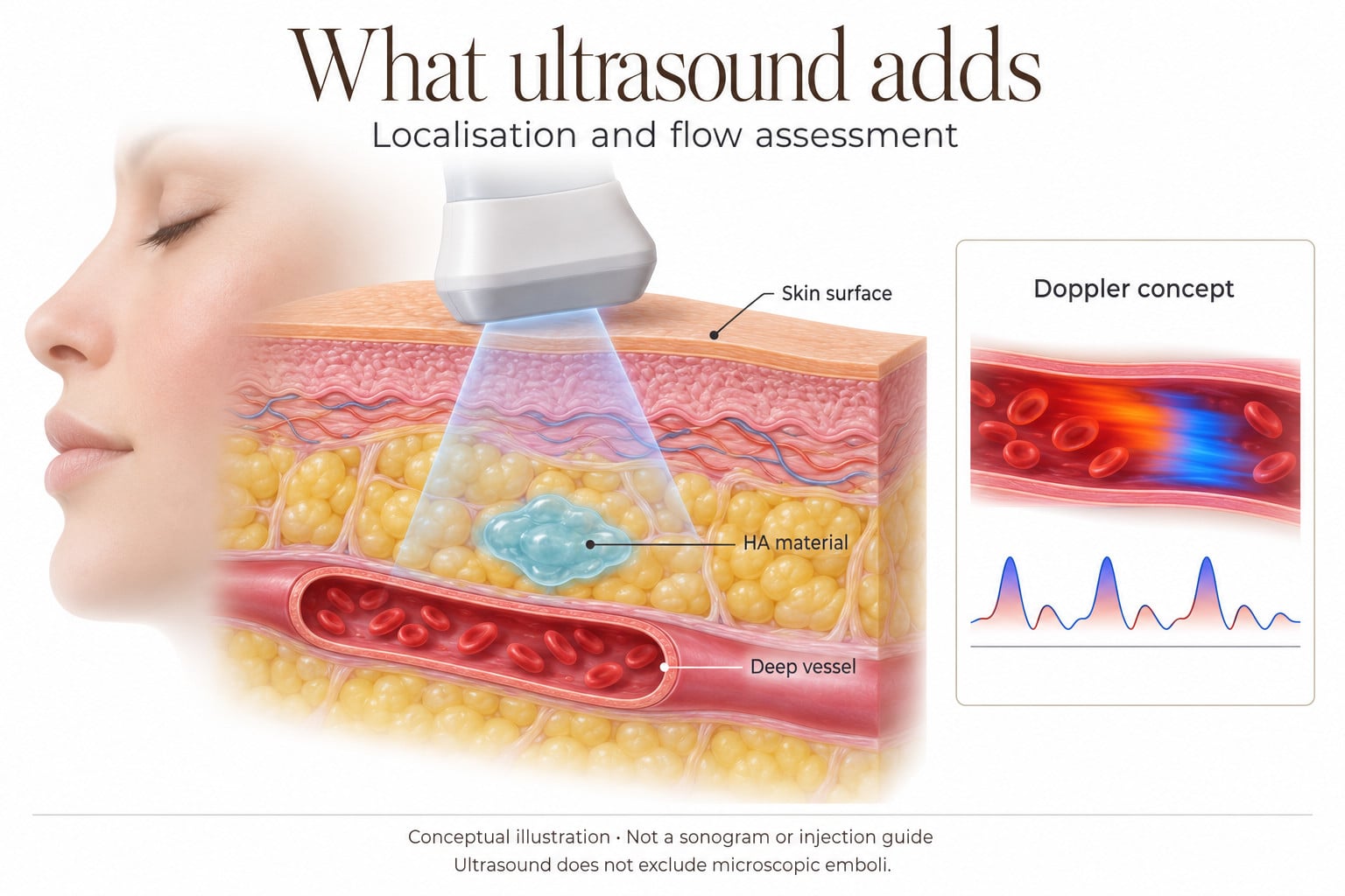
The most consequential shift of recent years is not a new drug but a new anatomical model, and it changes where the needle goes. Under Doppler ultrasound, targeted injection of a mean of 123 IU at the base of the main arterial trunk restored normal flow and resolved livedo and pain within 60 seconds in 100% of cases, in a series of 39 patients who had already failed conventional flooding with a mean of 1,519 IU and shown no or minimal improvement. All 39 healed without scar. The comparison within the same patients is what makes the series striking: the mean total dose fell from 1,519 IU to 122.5 IU, a statistically significant reduction, while the clinical response went from no improvement in 92.3% of patients to immediate improvement in all of them.
Pooled across four studies and 55 patients, ultrasound-guided hyaluronidase produced complete scar resolution in a probable 94.6%, against 77.8% for conventional non-image-guided injection. The mean dose used in the image-guided studies was 208 IU, a fraction of the 1500 IU conventional recommendation, and in two of the four studies ultrasound succeeded as salvage after conventional flooding had already failed. Targeting the filler deposit and injecting intra-arterially performed comparably, with complete resolution in 96.2% versus 83.3%, a difference that did not reach significance. Since HA deposits are far easier to see than small-calibre facial arteries, deposit-directed injection is also the more teachable technique; one study achieved clinical improvement with as little as 30 IU.
The ultrasound findings have since been extended beyond HA. A 2023 study characterized how CaHA appears sonographically depending on dilution and whether it is mixed with HA, which is the practical groundwork for identifying a non-HA deposit at the bedside rather than inferring its presence from the skin. An earlier paper made the case for early ultrasound in the diagnosis and treatment of vascular adverse events with HA fillers, and remains the origin of the targeted approach. The mechanistic logic is supported by the numbers in isolation: the plasma half-life of hyaluronidase is only two to three minutes, which is hard to reconcile with a claim that a superficial flood dissolves a millilitre of gel sitting deep in a perivascular space, whereas direct visual targeting of that mass resolves the clinical signs within a minute.
The caveats, stated plainly
Four studies, 55 patients, observational designs, and no randomised evidence possible when the alternative is withholding urgent treatment. Outcomes will be highly operator-dependent, and access to a clinician who can read a facial Doppler scan is not evenly distributed, which is why portable ultrasound has been slow to spread despite its obvious value. Intra-arterial work in small facial vessels realistically belongs to an interventional radiologist, and the intra-arterial subgroup in the pooled analysis rested on a single study of two patients. DeLorenzi flagged the underlying question that has still never been formally tested: when an artery is completely obstructed there is no flow past the blockage, so enzyme injected into the lumen tends to travel proximal to it and acts only where it touches the exposed surface, whereas flooding the whole zone appears to break down the filler along the vessel's entire course at once. Which is more effective remains open.
Bringing It Together: What To Do At The Chair
The literature converges on a triage model rather than a single universal first line. High-dose pulsed hyaluronidase should be given immediately, in every clinic, without waiting for anyone or any machine. Ultrasound then serves two purposes: as a salvage tool for cases that have not turned around within the first hour, and as a diagnostic instrument that tells you within minutes whether a presentation that looks mild on the surface is actually a stalled perforator sitting on a deep obstruction.
- Stop injecting and assess. Look for blanching, mottling, and slowed capillary refill, comparing against adjacent or contralateral normal skin; disproportionate pain is a finding in its own right.
- Treat immediately with high-dose pulsed hyaluronidase rather than watching. One dose given to a patient who turns out to have been fine costs nothing but the enzyme. An hour spent observing a livedo pattern costs tissue.
- Scale the dose to the ischemic surface area and repeat every 30 to 60 minutes until capillary refill, colour and pain normalise, with gentle massage after each injection.
- Use ultrasound as soon as it is available, targeting the perivascular space or the filler deposit rather than the skin that looks sick, and expect to need far less enzyme than the conventional protocol recommends.
- Refer for intra-arterial or retrobulbar intervention when the eye is involved or the response to initial treatment is inadequate. Ocular cases are a different emergency with a different window.
- Keep a written protocol, stock and expiry-check the enzyme, and know before the event whom you would call. Readiness, not dosage, is the variable most often missing when outcomes go badly.
If the picture is ambiguous, treat and reassess. The direction of travel is clear enough: smaller total enzyme doses, faster reperfusion, fewer scars, and a documented answer to the question every patient asks when something goes wrong, which is what you actually saw.
Source Publications
This review draws on the following fifteen publications.
Swipe or scroll horizontally to read the full references and source links.
| No. | Publication | Primary source |
|---|---|---|
| 1 | Soares DJ. Bridging a Century-Old Problem: The Pathophysiology and Molecular Mechanisms of HA Filler-Induced Vascular Occlusion (FIVO) – Implications for Therapeutic Interventions. Molecules, 2022; and Correction, Molecules, 2024. | Read source · Correction |
| 2 | Murray G, Convery C, Walker L, Davies E. Guideline for the management of hyaluronic acid filler-induced vascular occlusion. Journal of Clinical and Aesthetic Dermatology, 2021 (clinical staging as reproduced in the Soares review). | Read source |
| 3 | DeLorenzi C. New High Dose Pulsed Hyaluronidase Protocol for Hyaluronic Acid Filler Vascular Adverse Events. Aesthetic Surgery Journal, 2017. | Read source |
| 4 | Schelke LW, Velthuis PJ, Decates T, Kadouch J, Alfertshofer M, Frank K, Cotofana S. Ultrasound-Guided Targeted vs Regional Flooding: A Comparative Study for Improving the Clinical Outcome in Soft Tissue Filler Vascular Adverse Event Management. Aesthetic Surgery Journal, 2023. | Read source |
| 5 | Kadouch J, Schelke L, Groh O, Sokol V, Velthuis P. Intralesional hyaluronidase injection to relieve non-hyaluronic acid filler-induced vascular adverse events. International Journal of Dermatology, 2024. | Read source |
| 6 | Boey JJE, Boey JJJ, Chen Z, Cao T, Tan ASM, Ng ZY. Superior Outcomes with Ultrasound-Guided Hyaluronidase for Impending Filler-Induced Facial Skin Necrosis: A Systematic Review and Pilot Meta-Analysis. Aesthetic Plastic Surgery, 2025. | Read source |
| 7 | Boey JJJ, Boey JJE, Cao T, Ng ZY. Conventional High-Dose vs Low-Dose Hyaluronidase for Skin Necrosis after Hyaluronic Acid Fillers: A Systematic Review and Pilot Meta-Analysis. Aesthetic Plastic Surgery, 2024. | Read source |
| 8 | Alhusain AM, Alsaif M, Alkhathami AM, et al. Hyaluronidase Protocol in Management of Hyaluronic Acid Filler-Related Vascular Complications: A Systematic Review and Meta-Analysis. Aesthetic Plastic Surgery, 2026. | Read source |
| 9 | Borzabadi-Farahani A, Mosahebi A, Zargaran D. A Scoping Review of Hyaluronidase Use in Managing the Complications of Aesthetic Interventions. Aesthetic Plastic Surgery, 2024. | Read source |
| 10 | Alam M, Kakar R, Dover JS, Harikumar V, Kang BY, Wan HT, Poon E, Jones DH. Rates of Vascular Occlusion Associated With Using Needles vs Cannulas for Filler Injection. JAMA Dermatology, 2021. | Read source |
| 11 | Schelke LW, Velthuis PJ, Kadouch J, Swift A. Early ultrasound for diagnosis and treatment of vascular adverse events with hyaluronic acid fillers. Journal of the American Academy of Dermatology, 2023. | Read source |
| 12 | Ultrasonographic Patterns of Calcium Hydroxyapatite According to Dilution and Mix With Hyaluronic Acid. Journal of Ultrasound in Medicine, 2023. | Read source |
| 13 | Soares DJ, Trudel S, Siperstein R, Kean TJ, McCarthy AD. Microvascular Insights Into Hyaluronic Acid Filler Dispersal Within an Artificial Model of Arterial Embolism. Aesthetic Surgery Journal, 2026. | Read source |
| 14 | Murray G, Convery C, Walker L, Davies E. Guideline for the safe use of hyaluronidase in aesthetic medicine, including modified high-dose protocol. Journal of Clinical and Aesthetic Dermatology, 2021. | Read source |
| 15 | Walker L, Convery C, Davies E, Murray G, Croasdell B. Consensus opinion for the management of soft tissue filler induced vision loss. Journal of Clinical and Aesthetic Dermatology, 2021. | Read source |